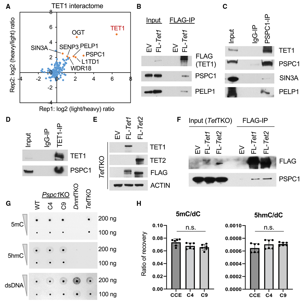
Figure 1. PSPC1 is an interacting partner of TET1 in ESCs.

(A) Protein ratios of FLAG-IP (TET1)versus Control-IP (empty vector) AP-MS in two replicates with reciprocal SILAC labeling are plotted, and a few proteins in the TET1 interactome are indicated.
(B and F) Co-immunoprecipitation (co-IP) of TET1 partners (B) or TET1/2 (F) by FLAG-IP followed by Western blot analysis in ESCs.
(C and D) Co-IP by endogenous PSPC1 (C) and TET1 (D) antibodies followed by western blot analysis in ESCs.
(E) Western blot analysis in Tet1/2/3 triple-KO (TetTKO) ESCs rescued with FLAG-tagged TET1 or TET2 in ESCs.
(G) DNA 5mC and 5hmC dot-blot analysis of WT and Pspc1KO (two independent clones, C4 and C9) ESCs. dsDNA antibody is reblotted as the loading control. Dnmt1/3a/3b triple-KO (DnmtTKO) and TetTKO ESCs serve as negative controls of 5mC and 5hmC, respectively.
(H) UHPLC-MS/MS quantification of 5′-methyl-deoxycytidine (5mC) and 5′-hydroxymethyl-deoxycytidine (5hmC) over deoxycytidine (dC) from genomic DNA of WT and Pspc1KO ESCs. Experiments were performed in biological duplicates with technical triplicates; p value is from two-tailed t test, and “n.s.” denotes statistically non-significant.
