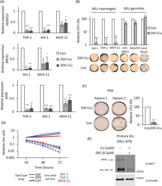
FIGURE 3.

Effects of MLL‐fusion depletion by disulfiram (DSF) treatment. (A) Effects of DSF–Cu on target gene expression in MLL‐rearranged cells. The expressions of HOXA10, MEIS1 and MYB were examined after 16 h with .3‐μM DSF or .6‐μM diethyldithiocarbamate (DDC) in the presence of 1 μM of Cu treatment through RT‐quantitative PCR (qPCR). The relative expression was normalized to 18 s. Bar charts are mean ± SD (t‐test, *p < .05, **p < .01, ***p < .001) from three independent experiments. (B) CFU assay of acute myeloid leukaemia (AML) or CD34+ cord blood cells treated with DMSO or .3‐μM DSF in the presence of 1 μM of Cu. Data represent the mean of three independent experiments. Bar charts are mean ± SD (C) CFU assay of two MLL‐rearranged patient–derived xenograft samples treated with DMSO or .3‐μM DSF in the presence of 1 μM of Cu. Data represent the mean of two independent patient samples repeated twice. Bar charts are mean ± SD and t‐test performed. (D) Anti‐proliferative activity of DSF and DDC against MLL‐rearranged AML. Cells were exposed to DMSO, DSF (.3 μM) or DDC (.6 μM) supplemented with 1 μM of Cu for periods varying from 0 to 72 h. Data illustrate a representative experiment of three independent replicates. Error bars are ±SD (E) western blot analysis of DSF effect on patient derived ALL MLL‐AF9 cells. MLL‐AF9 ALL cells were treated with .3‐μM DSF and 1‐μM Cu for 16 h. The proteins were detected using antibodies against N‐terminal MLL and vinculin
