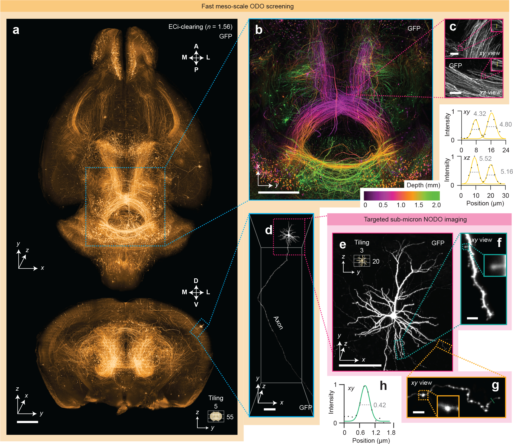
Figure 2 |. Fast meso-scale screening and targeted sub-micrometer imaging in cleared tissues.

(a) Fast meso-scale screening is performed of an entire intact ECi-cleared Slc17a7-Cre mouse brain with brain-wide axonal projections. (b) A depth-coded region of interest shows dense projections in the midbrain. (c) xy and xz zoom-in views illustrate the near-isotropic resolution of the hybrid OTLS microscope. Line profiles through individual axons demonstrate an ODO lateral and axial resolution of 4–5 μm at a large depth within the cleared specimen. (d-e) Targeted sub-micrometer imaging is performed of a region of interest around a cortical pyramidal neuron. (f-g) Zoom-ins of a dendrite and axon demonstrate sufficient lateral resolution to visualize individual spines and varicosities. (h) A line profile through an individual axon demonstrates a NODO lateral resolution of 0.42 μm within the cleared specimen. Scale-bar lengths are as follows: (a-b) 1 mm, (c) 10 μm, (d-e) 100 μm, and (f-g) 5 μm. All images are displayed without deconvolution. The imaging data in (a-g) was acquired from a single mouse brain in a single experiment.
