. 2022 Jun 2;13(24):7355–7364. doi: 10.1039/d2sc01769e

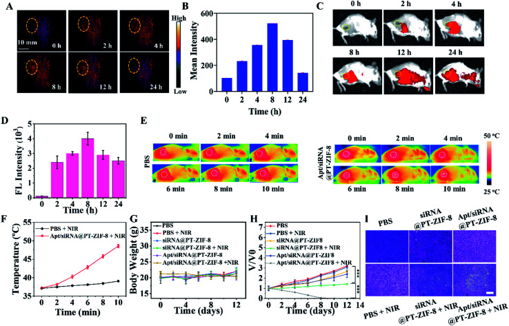
Fig. 5. (A, C) In vivo photoacoustic and fluorescence imaging of tumor-bearing mice after i.v. injection of Apt/siRNA@PT-ZIF-8 for 0, 2, 4, 8, 12, and 24 h. (B) The corresponding photoacoustic signal intensity in (A). (D) FI intensity of (C). (E) Thermal IR images of mice treated with PBS or Apt/siRNA@PT-ZIF-8 under 808 nm irradiation (2 W cm−2 for 2, 4, 6, 8, and 10 min). (F) Temperature changes on tumor sites according to the imaging in (E), PBS + NIR in the black line, and Apt/siRNA@PT-ZIF-8 + NIR in the red line. (G) Bodyweight monitoring during the process of (a) PBS, (b) PBS + NIR, (c) siRNA@PT-ZIF-8, (d) siRNA@PT-ZIF-8 + NIR, (e) Apt/siRNA@PT-ZIF-8, and (f) Apt/siRNA@PT-ZIF-8 + NIR groups. (H) Tumor volume change curves and all statistical data were represented as mean standard deviation (n = 4). (I) Representative H&E staining of the tumor slices collected from the mice on day 2. Scale bar: 100 μm.

Fig. 5