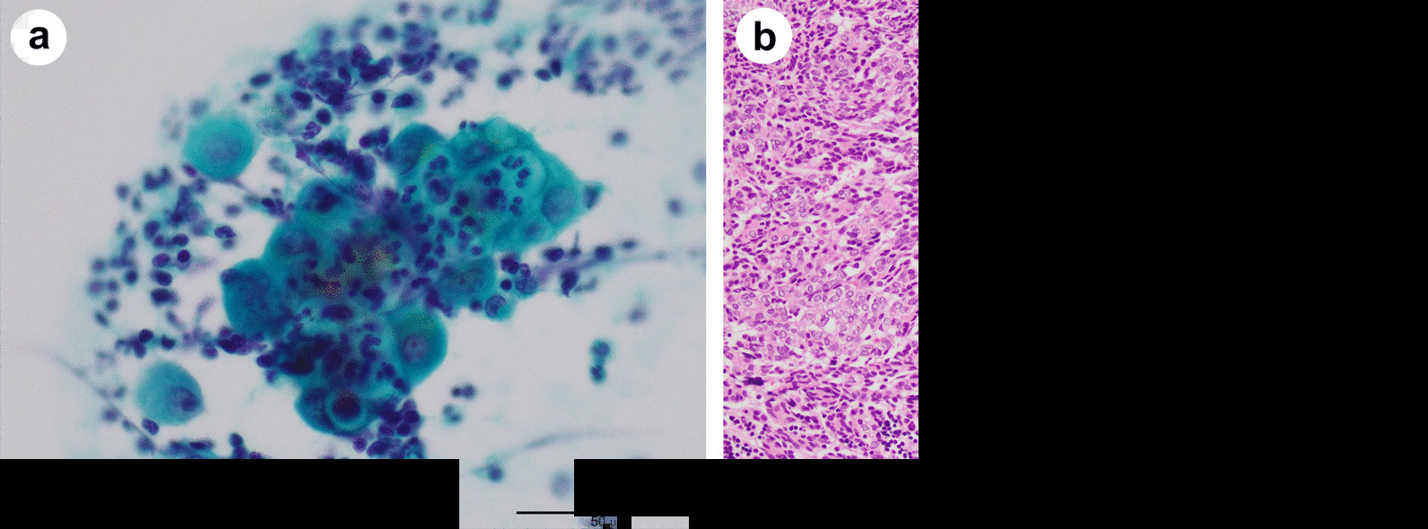
Fig. 2.

Cytology of pericardial fluid (Papanicolaou stain, × 400) (a) and histology of a biopsy specimen (Hematoxylin and eosin stain, × 200) (b). a Pericardial fluid showing an aggregate of atypical cells and inflammatory cells (mainly neutrophils). Atypical cells contain large nuclei with poor chromatin enrichment, relatively prominent nucleoli and abundant cytoplasm. b Spindle-shaped and oval epithelial cells with low-grade dysplasia arranged in pseudo-rosettes or short fascicles admixed with few lymphocytes. The sections were observed with a microscope: BX53 (OLYMPUS, Tokyo, Japan), lenses: UPlanFl (OLYMPUS, Tokyo, Japan), a camera: DP22 (OLYMPUS, Tokyo Japan), and a photo system: cellSens Standard 2.3 (OLYMPUS, Tokyo Japan) at a resolution of 96 dots per inch (1920 × 1440 pixels). The scale bar is 50 μm (a) or 100 μm (b)
