Figure 2.
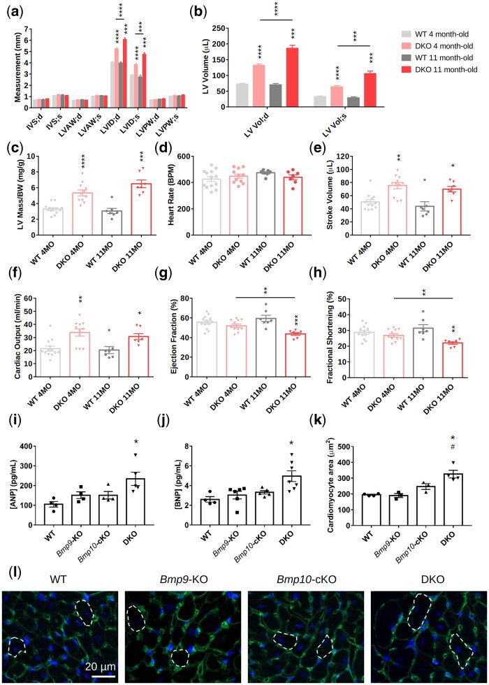
Combined loss of Bmp9 and Bmp10 leads to high cardiac output and cardiac hypertrophy in DKO mice. (A–H) Echocardiographic analysis of the left ventricle of 4- and 11-month-old WT and DKO female mice (n = 7–13/group). End-systolic (s) and end-diastolic (d) measurements of the interventricular septum (IV S), left ventricular anterior wall (LVAW), left ventricular posterior wall (LVPW) thickness, and the left ventricular internal diameter (LVID) (A), calculation of the left ventricular volume (LV Vol) (B), left ventricular mass/body weight (LV mass/BW) (C), heart rate (D), stroke volume (E), cardiac output (F), ejection fraction (G), and fractional shortening (H). All mice were injected with tamoxifen at the age of 2 months. Data are presented as the mean ± SEM and were analysed using Mann–Whitney tests to compare DKO vs. WT or 4-month-old vs. 11-month-old mice. *P < 0.05, **P < 0.01, ***P < 0.001, ****P < 0.0001. (I–L) Plasma analysis and immunofluorescence. ANP (atrial natriuretic peptide) (I) and BNP (brain natriuretic peptide) (J) plasma levels (n = 4–7 male mice/group). Quantitative analysis of cardiomyocyte size (K) and representative photomicrographs (L) of transverse heart sections stained with FITC-conjugated wheatgerm agglutinin to outline individual cardiomyocytes (n = 3–4 female mice/group, 100 cardiomyocytes/mouse). Mice were injected with tamoxifen at the age of 2 months and analysed at the age of 4–5 months. Data are presented as the means ± SEM and were analysed using Kruskal–Wallis tests followed by Dunn’s tests. *P < 0.05 vs. WT; #P < 0.05 vs. Bmp9-KO.

