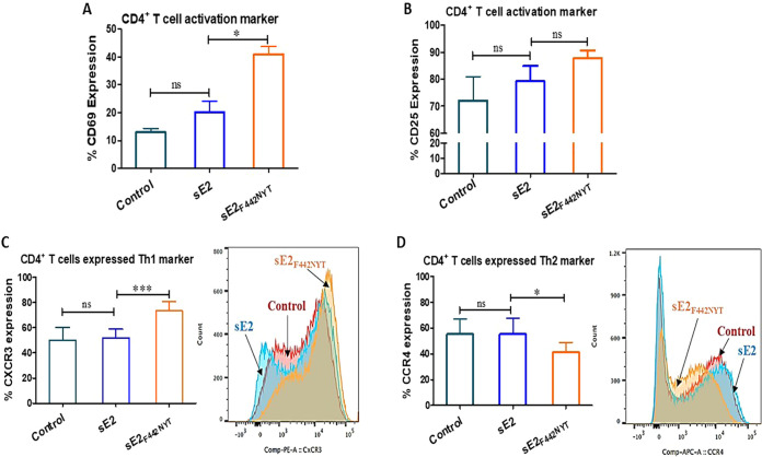
FIG 3.
Effect of macrophage activation by sE2F442NYT on the polarization of CD4+ T cells toward a Th1 type. (A and B) CD4+ T cells were isolated from healthy donors (n = 4) and treated with sE2 or sE2F442NYT for 2 h in the presence of human anti-CD3 (5 μg/mL) and anti-CD28 (1 μg/mL). The expression of T cell activation markers CD69 and CD25 on the surfaces of CD4+ T cells was quantified by flow cytometry. (C and D) Monocyte-derived macrophages isolated from healthy donors (n = 6) were treated with sE2 or sE2F442NYT protein for 24 h, and autologous CD4+ T cells were cocultured with the treated macrophages for 4 days. Th1 and Th2 polarization was measured as the expression of CXCR3 and CCR4, respectively, on the surfaces of CD4+ T cells by flow cytometry. The significance level is indicated (*, P < 0.05; ***, P < 0.001; ns, not significant).

