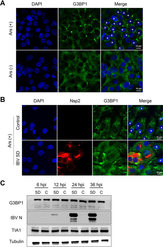
FIG 2.
IBV replication abrogated eukaryotic translation initiation factor 2α (eIF2α)-dependent SG formation. (A) CEK cells were treated with 1 mM sodium arsenite for 30 min and then immunostained. (B) CEK cells were infected with IBV SD at 18 hpi and then with 1 mM sodium arsenite for 30 min and then immunostained. Cells were detected with anti-Nsp2 antibody (red), SGs with anti-G3BP1 (green), and cell nuclei with DAPI (blue). Representative images of three independent experiments are shown. Scale bars = 10 μm. (C) CEK cells were mock infected (control [C]) or infected with 106.0 50% egg infective dose of IBV rSD. At 6, 12, 24, and 36 hpi, cell lysates (10 μg per lane) were analyzed via Western blot to detect G3BP1, IBV N, TIA1, and tubulin. Small asterisks indicate SG formation in the cells.

