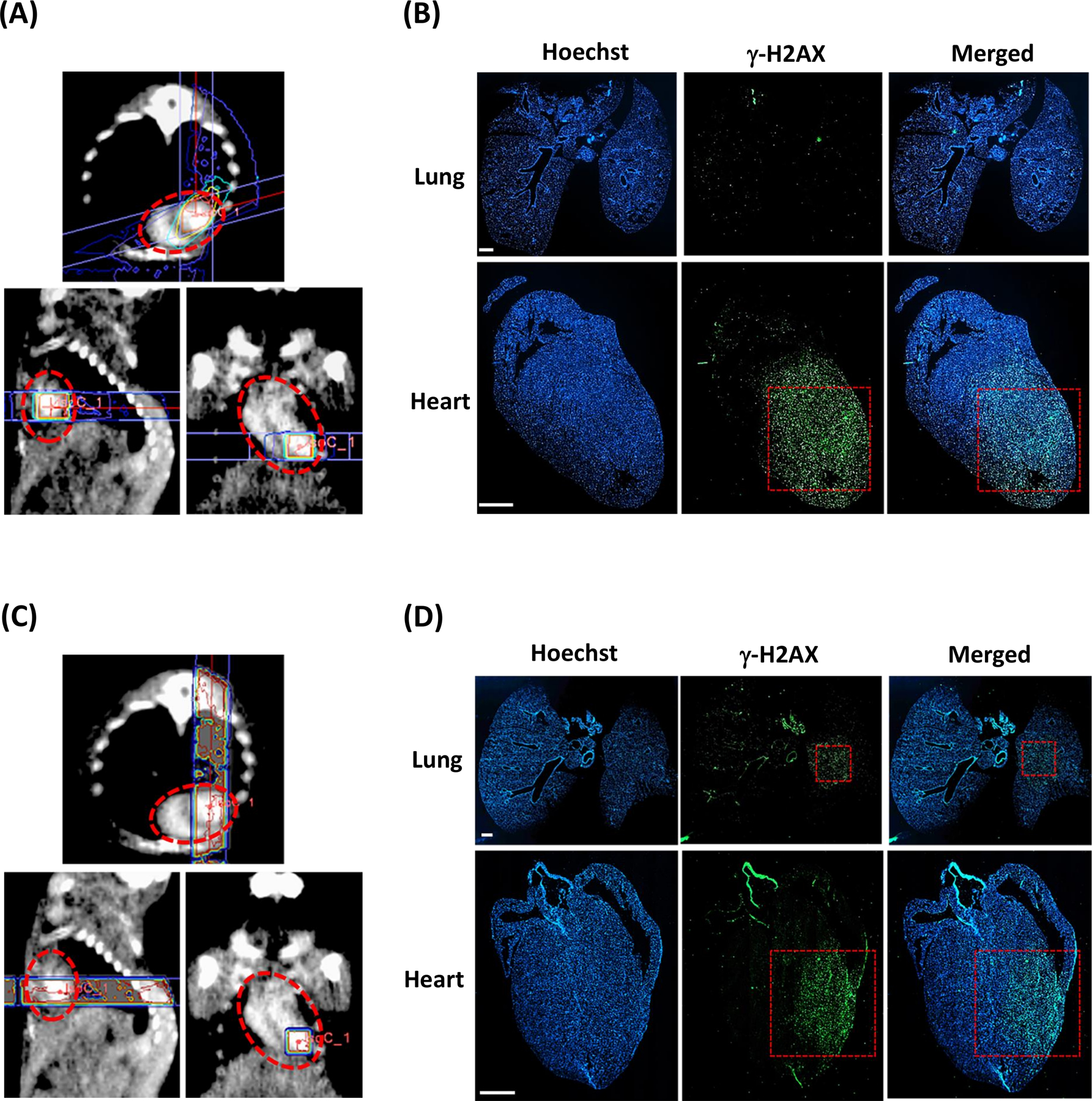
Figure 2. Dose plan in MuriPlan and corresponding immunohistochemical γ-H2AX staining of heart and lung tissue.

A, Axial, sagittal, and frontal views of RT planning and delivery to the cardiac apex at a dose of 60 Gy with a 3×3mm collimator and conformal RT over a 75° arc. B, Positive staining in the cardiac apex and negative staining in the lungs confirms selective PH RT targeting. C, Axial, sagittal, and frontal views of RT planning and delivery to the cardiac apex at a dose of 40 Gy using two 20 Gy 3×3mm vertical opposing beams. D, Positive staining in both the cardiac apex and in the mid-left lung confirms PHL RT targeting. The red dotted circles outline the cardiac silhouette. The red dotted boxes indicate the 3×3mm collimator. Scale bar 1mm.
