
Figure 2. TSST-1 IgG does not prevent TSST-1+ Staphylococcus aureus colonization of BP lesions.
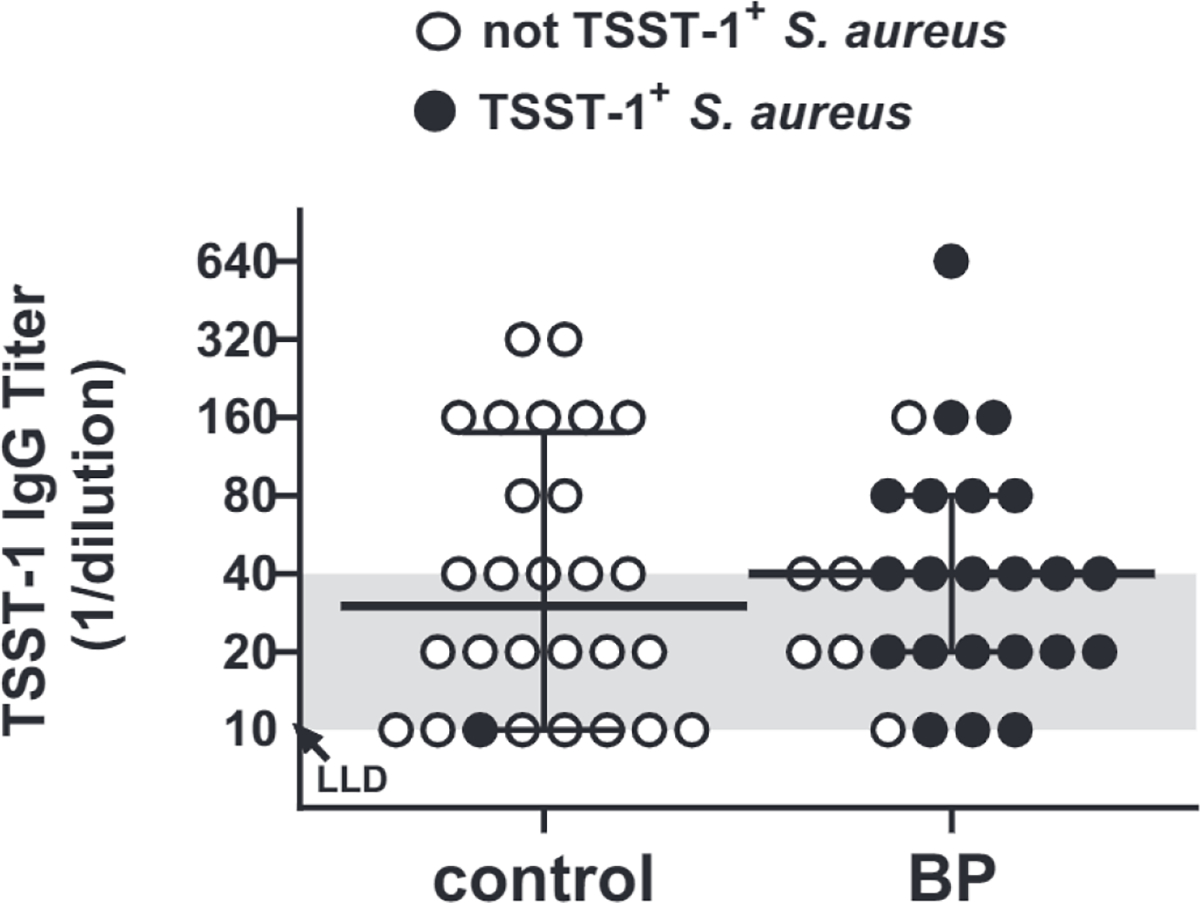
Serum titers of TSST-1–specific IgG were measured by ELISA. Titers ≤40 (shaded area) are not considered protective. TSST-1 IgG titers were similar in the sera from 28 patients with BP and 28 matched controls (Mann–Whitney test, P = 0.787), with 50% and 57% of each respective group showing protective titers. TSST-1+ S. aurous colonization (filled dot) was not correlated with the level of TSST-1 IgG (Spearman’s r2 = 0.005, P = 0.9796). Each point represents the average of triplicate wells for an individual patient, with the median indicated by the solid line. BP, bullous pemphigoid; LLD, lower limit of detection; TSST-1, toxic shock syndrome toxin-1.