FIGURE 3.
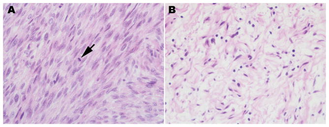
Cellular composition of neurofibromas. A, hematoxylin and eosin stained section of a dermal neurofibroma (40x). All subsequent images are taken from this same tumor at a 63x magnification. B, Immunoreactivity for the transcription factor Sox10 (green, representative cell indicated by an arrow) in the Schwann cell component of this dermal neurofibroma. The section has been counterstained with bisbenzamide to highlight the nuclei of all cells in the tumor. C, Immunoreactivity for the mast cell marker CD117 (also known as c-Kit; red, representative cell indicated by arrow) in the dermal neurofibroma. D, Immunoreactivity for CD34 (red) is apparent in vasculature (arrow) and in small dendritic cells in the tumor. E, Immunoreactivity (red) for the fibroblast nuclear marker TCF4 (transcription factor 4; representative cell indicated by arrow) in the tumor. F, Immunoreactivity for the pan-macrophage marker Iba1 (red, representative cell indicated by arrow) in the tumor. G, Immunoreactivity for CD163 (red), a marker of the M2 (anti-inflammatory) subclass of macrophages, in the tumor (representative cell indicated by arrow). H, Immunoreactivity for CD86 (red), a marker of the M1 (pro-inflammatory) subclass of macrophages, in the tumor (representative cell indicated by arrow).

