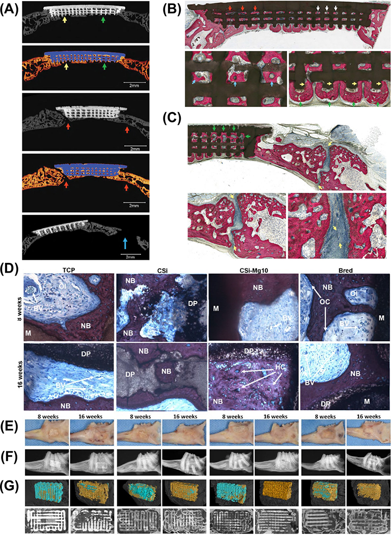
Figure 18.
(a) Micro–computed tomographic slices of new bone formation within scaffold at 8 weeks. The osseoconductive infiltration through the scaffolds (green and yellow arrows on second row) and bone regeneration in immature skeletal calvaria did not affect the suture patency (red arrows). (b-c) Histological examination showing bone formation is guided by highly osseoconductive scaffold dimensions as new bone formation is directed from scaffold pore-to-pore while interacting with scaffold struts. Adapted from.177 (d) Histological analysis of bone defect site in rabbit mandibles, ×20 (e) Images of the specimens. (f) Radiographs of the specimens. (g) Two-dimensional, 3-dimensional micro–computed tomography (CT) images of the scaffolds implanted in the rabbit alveolar defects with different time separately. Adapted from.126

