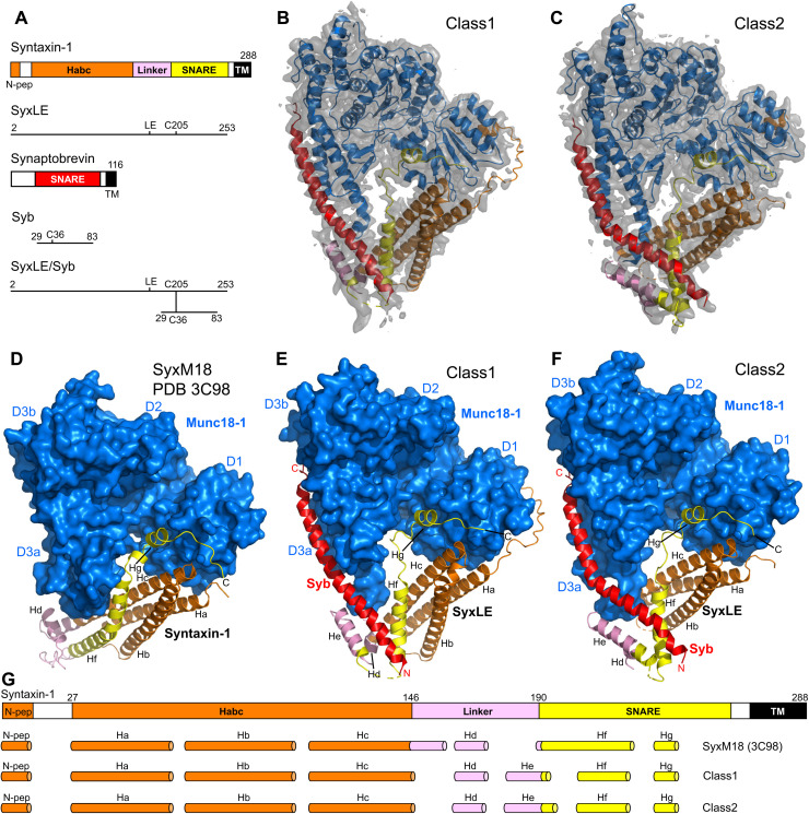
Fig. 1. Two cryo-EM structures of the template complex.
(A) Domain diagrams of syntaxin-1 and synaptobrevin, and summary of the fragments used to prepare SyxLE/Syb. N-pep, N-peptide; SNARE, SNARE motif. (B and C) 3D reconstructions of two structures of the template complex, class1 (A) and class2 (B), and corresponding ribbon diagrams fitted into the cryo-EM maps at 3.7 and 3.5 Å, respectively. (D to F) Comparison of the crystal structure of the syntaxin-1–Munc18-1 complex (SyxM18) (PDB code 3C98) (8, 34) (D) with the two cryo-EM structures of the template complex, class1 (E) and class2 (F). The surface of Munc18-1 is shown in blue, and the SNAREs are represented by ribbon diagrams with synaptobrevin (Syb) in red and syntaxin-1 in orange (N-peptide and Habc domain), pink (linker), and yellow (SNARE motif). The domains of Munc18-1 (D1, D2, D3a, and D3b) are labeled. The helices formed by syntaxin-1 (named Ha-Hg) are indicated. The N and C termini of Syb, as well as the C terminus of SyxLE, are labeled. (G) Summary of the locations of the helices observed in SyxM18, class1, and class2. The helices are represented by cylinders, and they are shown below a domain diagram of syntaxin-1 with selected residue numbers above to indicate domain boundaries. The helix formed by the N-peptide at the very N terminus is named N-pep, and subsequent helices of class1 and class2 are named Ha to Hg. To facilitate comparisons, the same nomenclature is used for SyxM18 helices in the same or similar positions as those observed in class1 and class2, but not that there is no He helix in SyxM18.

