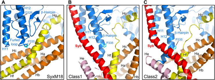
Fig. 2. Structural changes in Munc18-1 that lead to synaptobrevin binding and template complex formation.
(A to C) Close-up views of the area where the Munc18-1 loop unfurls to allow synaptobrevin binding in the syntaxin-1–Munc18-1 complex (SyxM18) before unfurling (A) and in class1 (B) and class2 (C), where the loop is unfurled and synaptobrevin is bound. Munc18-1 is colored in blue, synaptobrevin (Syb) in red, and syntaxin-1 in orange (N-peptide and Habc domain), pink (linker), and yellow (SNARE motif). P335 is shown as a stick model, with carbon atoms in cyan. The positions of the furled loop and D326 in the Munc18-1–syntaxin-1 complex, as well as of synaptobrevin (Syb), P335, a nearby β-hairpin, and selected helices of Munc18-1 and syntaxin-1 are indicated.

