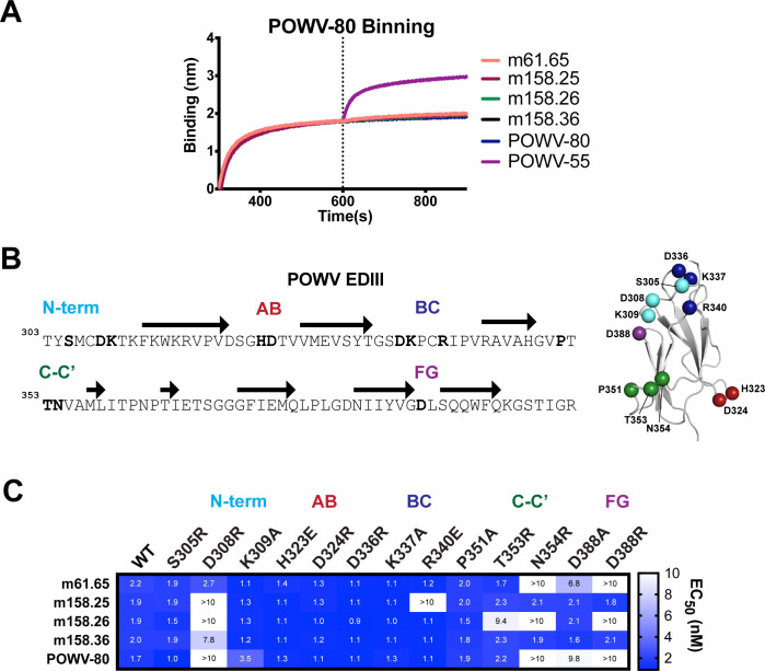
Fig 6. Epitope binning and mutagenesis mapping of neutralizing POWV EDIII mAbs.
(A) Two-phase binding of neutralizing mAbs to POWV EDIII against mAb POWV-80 by BLI. MAb POWV-80 was bound to a sensor loaded with POWV EDIII followed by the sequential addition of the indicated second mAb. Representative data from two independent experiments is shown. (B) Sequence map of POWV EDIII for mutagenesis studies. Bolded residues in N-terminus, AB, BC, C-C’ and FG loops indicate positions where substitutions were introduced. Structure of POWV EDIII with mutations highlighted as Cα spheres is shown. (C) Heatmap of EC50 values of mAbs against MBP-POWV-EDIII mutants. EC50 values were calculated using non-linear regression analysis of ELISA binding curves performed twice independently in triplicate wells (mean ± SD).

