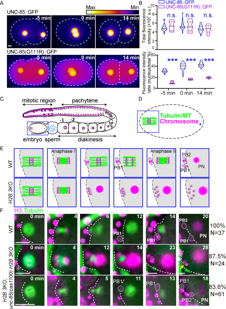
Fig 2. UNC-85 mutations restore the meiosis defects of H2B 3KO animals.
(A) Fluorescence time-lapse heatmaps of UNC-85::GFP (up) and UNC-85::GFPG111R (down) in one-cell stage embryos. The first occurrence of pronucleus meeting was defined as time 0 min. Scale bar, 5 μm. (B) Quantifications of the whole-cell GFP-tagged UNC-85 fluorescence intensity (up) and intensity ratio of the nucleus to the cytoplasm (down). Data are presented as mean ± SD (error bars), N = 11–34. Statistical significance is based on two-way ANOVA, n.s., not significant, ***p< 0.001. (C) Schematic representation of an adult hermaphrodite gonad. (D) Schematic depiction of the fertilized embryo. MTs and chromosomes are indicated by green and magenta, respectively. (E) Enlarged schematic timescales of oocyte meiosis in WT (up) and H2B 3KO (down). (F) In utero fluorescence time-lapse images of meiosis visualized by GFP::tubulin (green) and mCherry::Histone (magenta) during oocyte meiosis in WT, H2B 3KO and H2B 3KO; unc-85(cas1100) mutant. Metaphase Ӏ was time zero. N (N = 24–61 animals, oocytes are from different animals) and and percentages of meiosis patterns were indicated on the right. Scale bar, 5 μm.

