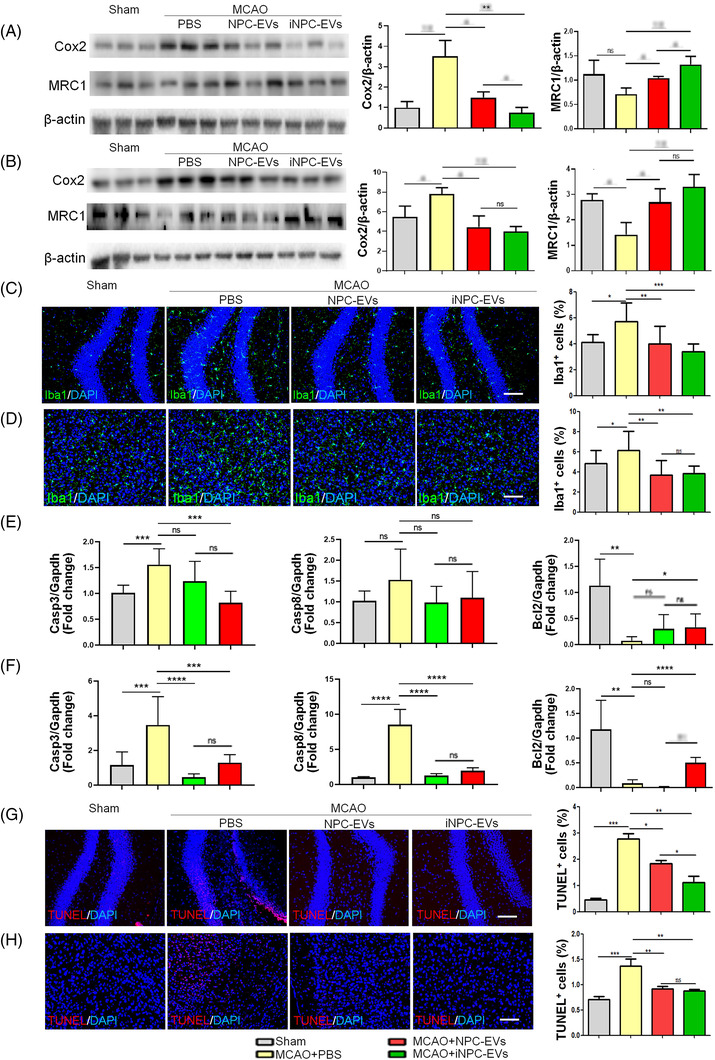
FIGURE 3.

Post‐stroke administration of induced neural stem/progenitor cell (iNPC)‐extracellular vesicles (EVs) inhibits neuroinflammation. Focal cerebral ischemic brains treated with or without EVs, and their sham controls were collected on day 28 after middle cerebral artery occlusion (MCAO). (A and B) Representative blot (left) and quantification (right) of Cox2 and MRC1 protein expression levels in the hippocampus (A) and the peri‐infarct cortex (B) (n = 3). (C and D) Representative confocal microscopy images of Iba1 immunostaining (green) in the hippocampus (C) and the peri‐infarct cortex (D). Proportions of cells with Iba1 immunoreactivities were given on the right panel (n = 4). (E and F) Transcript levels of Casp3, Casp8 and Bcl2 in the hippocampus (E) and the peri‐infarct cortex (F) were determined by quantitative reverse transcription polymerase chain reaction (RT‐qPCR) analyses (n = 4). (G and H) Representative confocal microscopy images of TUNEL (red) staining in the hippocampus (G) and the peri‐infarct cortex (H). Proportions of TUNEL positive cells were given on the right panel (n = 4). RT‐qPCR data were normalized to Gapdh and presented as fold changes compared with sham control groups. Western blotting data were normalized to β‐actin. Scale bar: 100 μm. Error bars denote s.d.. *p < .05, **p < .01, ***p < .001, and ****p < .0001. ns denotes non‐significance. The statistical difference among groups was assessed with the parametric one‐way ANOVA with post hoc Bonferroni test
