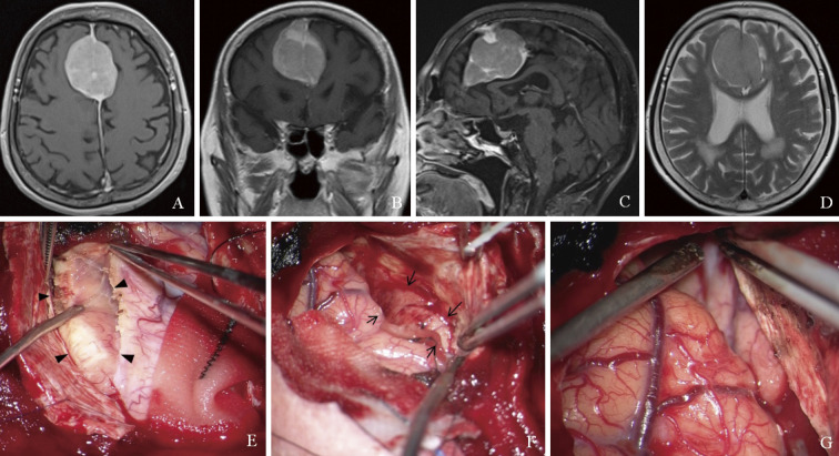
Fig. 1.
Magnetic resonance imaging shows a solitary mass lesion at the cerebral falx with contrast enhancement on T1-weighted imaging (A: axial, B: coronal, and C: sagittal views) and no apparent findings of invasion and edema around the brain on T2-weighted imaging (D). Intraoperative photographs show that the lesion is elastic soft tissue. First, the right side of the lesion is removed (E, arrowheads), and next the left side is also removed (F, arrows). Finally, the dura mater attachment of the mass is cut and removed (G).

