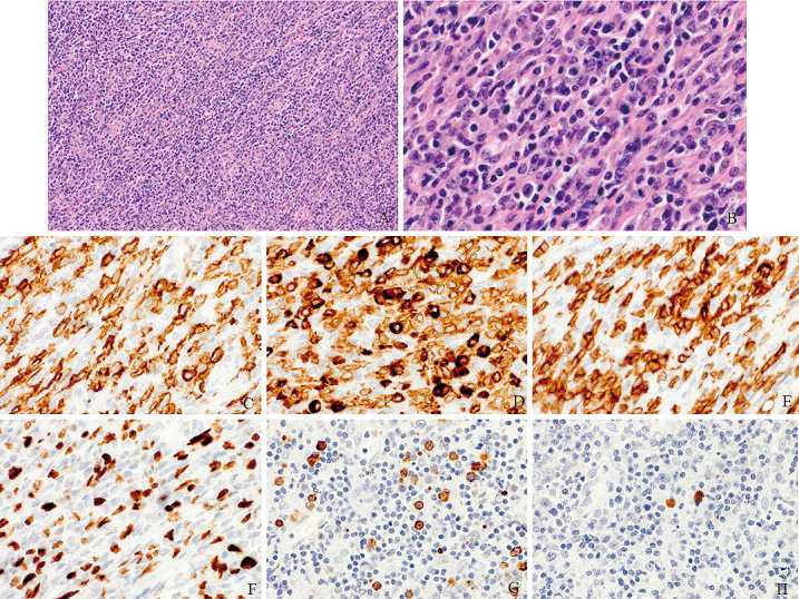
Fig. 2.

Surgical specimen stained with hematoxylin-eosin shows diffuse infiltration of small lymphocytes and plasma cells. There is some proliferation of large lymphocytes with folded nuclei, high-density chromatin, and inconspicuous nucleoli (A: ×100 and B: ×400). Immunohistochemical examinations show that these cells exhibit positive staining for CD20 (C) and CD79a (D), and there are CD3-positive T-cells (E). The atypical lymphocytes are stained for mind bomb 1 (F). The plasma cells are positive for the lambda immunoglobulin light chain (G) but lack kappa light chain expression (H).
