
Fig. 3.
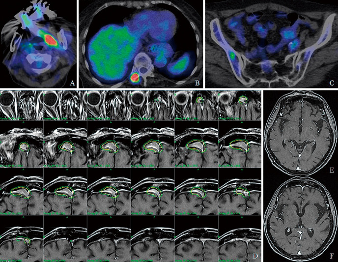
18F fluoro-2-deoxyglucose positron emission tomography shows high accumulations at the left pterygoid muscle (A) and the right transverse processes of the thoracic vertebrae (B), and mild accumulation at the right ilium bone (C). The plan of gamma knife surgery shows a maximum dose of 30 Gy and a marginal dose of 15 Gy (yellow lines) for thickening of the dura mater with contrast enhancement (D). Magnetic resonance images two years later show no evidence of recurrence (E and F).