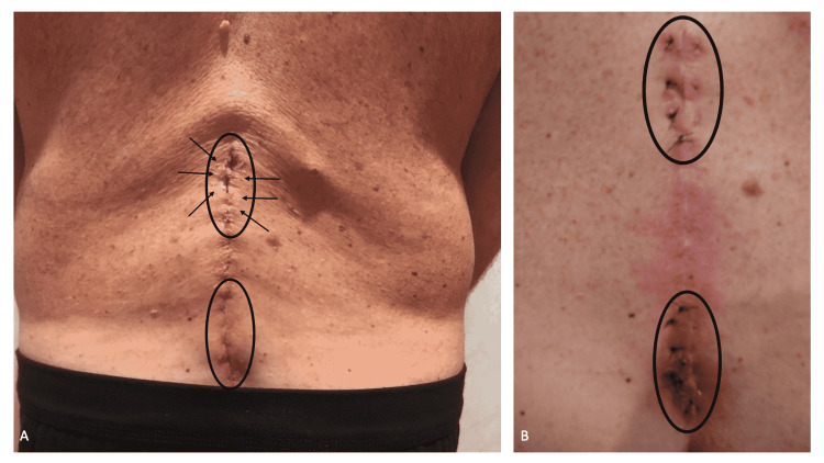
Figure 1. Clinical presentation of the cerebrospinal fluid leak draining onto the back skin and suture repair of the duro-cutaneous fistulas.
The lower back of a 58-year-old man, who had back surgery 12 days earlier, shows the sites where cerebrospinal fluid has leaked onto the skin (black ovals); superiorly (upper black oval), flakes of dried cerebrospinal fluid (black arrows) can be observed (A). Subsequently, sutures were placed deeply into the subcutaneous tissue to compress the area affected by the duro-cutaneous fistulas and thereby prevent any further leakage of cerebrospinal fluid to the skin’s surface (B).

