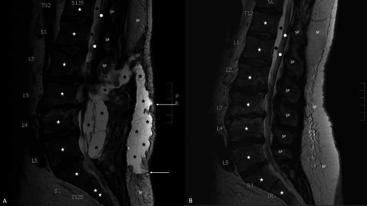
Figure 2. Post-surgical and pre-surgical sagittal views of magnetic resonance imaging with contrast of the lower back.
A magnetic resonance imaging of the lumber back of a 58-year-old man was performed on a 1.5 Tesla scanner, with sagittal short T1 inversion recovery, T1-weighted, and T2-weighted imaging 12 days after surgery to evaluated him for a post-operative cerebrospinal fluid leak (A). The sagittal view demonstrates the vertebral bodies (white stars) and spinal process (labeled SP) of the lower thoracic (labeled T12), lumbar (labeled L1 to L5) and upper sacral (labeled S1) vertebrae. The lower portion of the spinal cord (white dots) and cerebrospinal fluid in the dura (black dots) can also be seen. In addition, subcutaneous fat (labeled SF) can be noted. There are two large, connected collections of cerebrospinal fluid (black stars) in the soft tissue of the back; two tracts containing cerebrospinal fluid extend from the soft tissue collection to the skin surface (white arrows). The sagittal view of a similarly performed magnetic resonance imaging, from 18 months earlier, does not show any leakage of cerebrospinal fluid (B).

