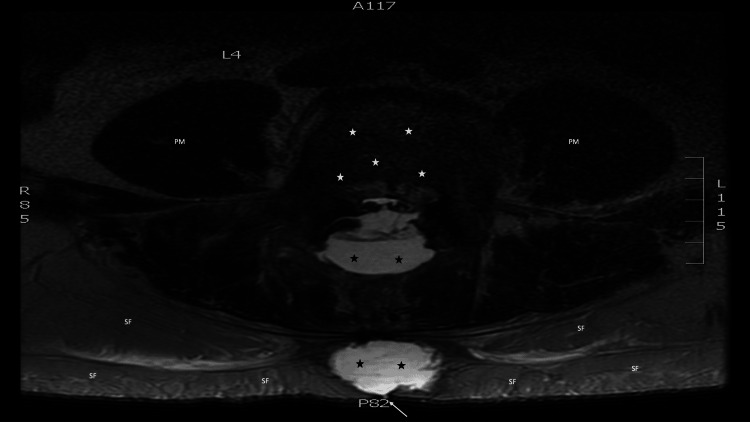
Figure 3. Axial view of magnetic resonance imaging showing the cerebrospinal fluid leak.
Twelve days after surgery, a magnetic resonance imaging of the lumber back of a 58-year-old man was performed on a 1.5 Tesla scanner, with axial T1-weighted and T2-weighted imaging. The axial view, at the level of the fourth lumbar vertebrae, demonstrates the vertebral body (white stars) and bilateral psoas muscles (labeled PM). Subcutaneous fat (labeled SF) can be seen. In addition, two collections of cerebrospinal fluid (black stars) are present in the soft tissue of the back; a tract (white arrow), extends from one of the soft tissue collections of cerebrospinal fluid to the surface of the skin surface.

