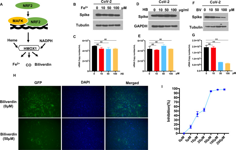
Fig. 2.
HMOX1 product biliverdin suppresses SARS-CoV-2 replication. A A schematic diagram showing the heme oxygenase pathway. Vero-E6 cells were infected with recombinant GFP-SARS-CoV-2 at an MOI of 0.01. After incubation with virus for 1 h, the cells were treated with the indicated doses of Fe2+ (B) hemoglobin (HB) D or biliverdin (F) for 24 h. C, E, G The amount of viral RNA in the cell culture medium was quantified by real-time PCR assay. H Fluorescence microscopy images of GFP-SARS-CoV-2 in biliverdin-treated ACE2-HeLa cells. Blue: DAPI (nuclear staining). I The dose response curve of biliverdin against SARS-CoV-2. HeLa-ACE2 cells were infected with recombinant GFP-SARS-CoV-2. After incubation with virus for 1 h, the cells were treated with biliverdin at different concentrations for an additional 24 h. The viral NP expression in the SARS-CoV-2-infected cells was quantified by real-time PCR and normalized to the expression of the 36B4 gene. The results are expressed as the mean ± SD (error bar) of 3 independent experiments; asterisks represent statistical significance based on two-tailed unpaired Student’s t test (*P < 0.05, **P < 0.01)

