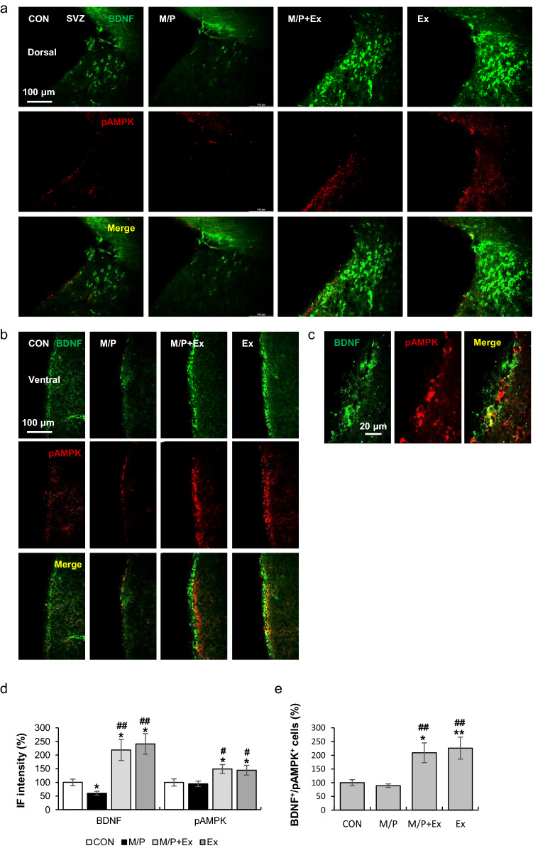
Figure 3.
Rotarod walking exercise activated the AMPK/BDNF pathway in the SVZ area of M/P-treated mice. (a) Photomicrographs of the double fluorescent staining of dorsal SVZ BDNF+ and pAMPK+ cells (CON, N = 8; M/P, N = 9; M/P + Ex, N = 9; Ex, N = 8, 200 × of magnification). (b) Photomicrographs of the double fluorescent staining of ventral SVZ BDNF+ and pAMPK+ cells (CON, N = 8; M/P, N = 9; M/P + Ex, N = 8; Ex, N = 9, 200 × of magnification). (c) Representative photomicrograph of the high magnitude of SVZ BDNF+/pAMPK+ cells (400 × of magnification). (d) Quantification of the fluorescent intensity of SVZ BDNF+ and pAMPK+ particles showing that exercise restored the M/P-induced decrease in BDNF+, and enhanced the pAMPK+ IF intensity, regardless of MPTP treatment (CON, N = 8; M/P, N = 9; M/P + Ex, N = 9; Ex, N = 8). (e) Quantification of the fluorescent staining of SVZ BDNF+/pAMPK+ cells showing that exercise increased the number of BDNF+/pAMPK+ cells, regardless of MPTP treatment. Data are presented as mean ± SEM. *p < 0.05, vs. CON; **p < 0.01, vs. CON; #p < 0.05, vs. M/P; ##p < 0.01, vs. M/P.

