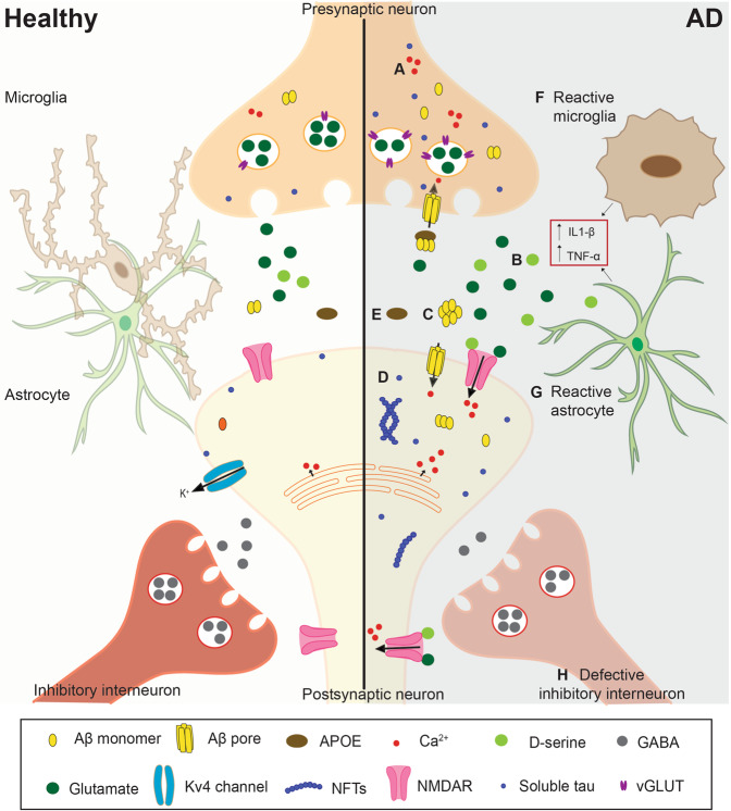
Fig. 1. Mechanisms causing neuronal hyperexcitability in Alzheimer’s disease.
Representation of a synapse in the healthy (left) and in the AD (right) brain. A Increased release of calcium from intracellular stores from pre and postsynaptic neurons results in higher levels of cytosolic calcium. B Enhanced glutamatergic signaling can be caused by reduced astrocytic uptake, reduced levels of glutamine synthetase (not shown in the Figure), and/or increased vGLUT expression. C Amyloid-β can form ionic pores in the plasma membrane. It also reduces the expression of Kv4 channels and increases NMDAR activation via increased d-serine and glutamate release and reduced glutamate uptake. D Protein tau can contribute to hyperexcitability by altering glutamate levels as well as the expression and function of Kv4.2 channels and NMDAR. E Compared to apoE3, apoE4 reduces the clearance and uptake of Aβ42 by astrocytes and microglia, respectively. F, G The release of pro-inflammatory cytokines, such as IL-1β and TNF-α, by glial cells can promote hyperexcitability. F During gliosis, reactive microglia fail to properly regulate neuronal excitability. G In AD, astrocytes show increased release and reduced uptake of glutamate, as well as reduced expression of potassium channel Kir4.1 (not shown in the Figure). Reactive astrocytes also increase neuronal excitability by reducing synaptic inhibition. H Reduced firing frequency and the number of inhibitory GABAergic neurons is another contributing factor to hyperexcitability in AD. AD Alzheimer’s disease, NMDAR NMDA receptor, vGLUT vesicular glutamate transporter.

