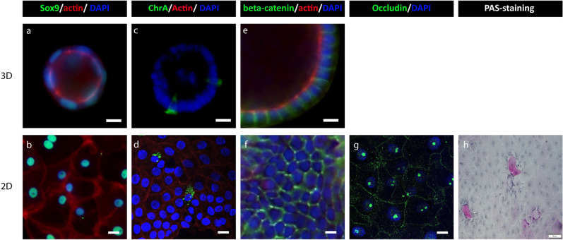
Figure 6.
Identification of different cell structures in 3D chicken intestinal organoid and the monolayers. Immunohistochemistry images of 3D and 2D structures of chicken intestinal organoids, processed 2 days after seeding. (a,b) Progenitor cells are visualized with anti-Sox9. (c,d) Enteroendocrine cells are visualized by Chromogranin A. (e–g) Tight junctions are visualized with beta-catenin or occludin (h) Mucus-containing goblet cells are visualized using Periodic Acid Schiff (PAS)-positive cell staining of 2D chicken intestinal organoids. The actin filaments are visualized with rhodamine phalloidin (red), and nuclei are stained with DAPI (blue) Scale bar: 400 µm.

