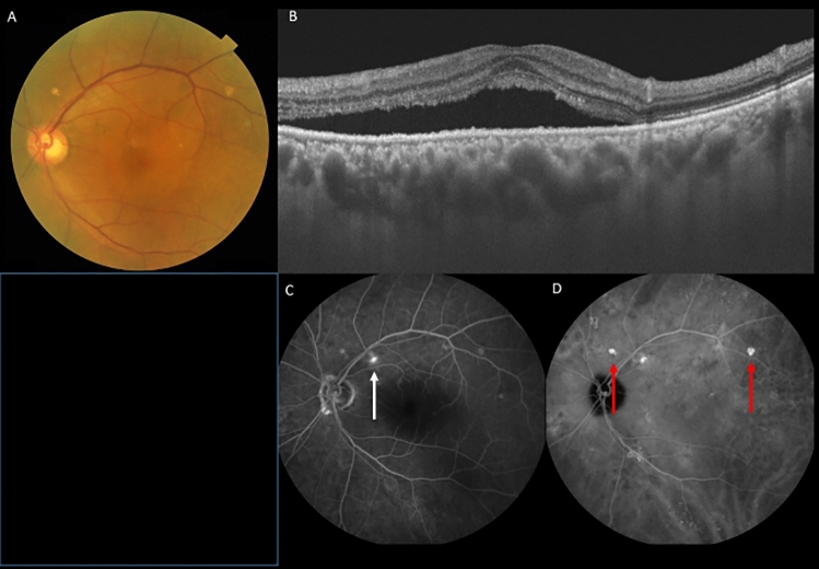
Figure 4.
A 70-year-old male patient with central serous chorioretinopathy and pachydrusen in the left eye. (A) Fundus photography shows pachydrusen outside the arcade and serous retinal detachment in the macula in the left eye. (B) Swept-source optical coherence tomography demonstrated serous retinal detachment and a dilated choroid in the left eye. (C) Fluorescein angiography revealed the leakage point indicating by a white arrow in the left eye. (D) Late phase indocyanine green angiography revealed hyperfluorescent areas corresponding to pachydrusen indicating by red arrows in the left eye.

