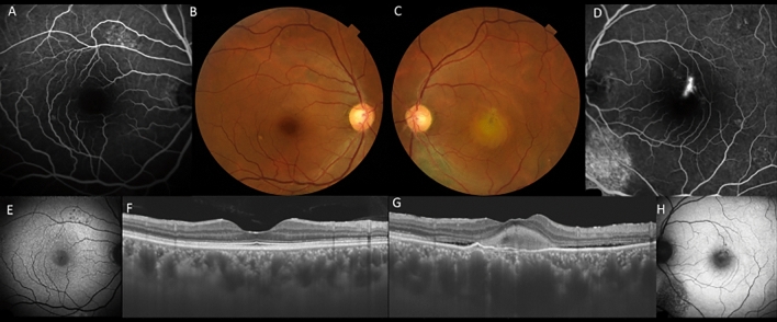
Figure 5.
A 48-year-old male patient with central serous chorioretinopathy and fibrin in the left eye. (A) Retinal pigment epithelial changes were seen superior to the macula by fluorescein angiography (FA). (B) There were no obvious abnormal findings in the right eye. (C) A white round lesion was seen along with retinal pigment epithelial atrophy inferior to the optic disc in the left eye. (D) A pinpoint leakage and retinal pigment epithelial atrophy inferior to the optic disc was seen by FA in the left eye. (E) Fundus autofluorescence (FAF) shows hypofluorescent spots corresponding to retinal pigment epithelial changes in FA in the right eye. (F) Swept-source optical coherence tomography (SS-OCT) shows dilated choroid without exudation in the right eye. (G) SS-OCT shows hyperreflective materials with shallow serous retinal detachment in the left eye. (H) FAF shows hyperautofluorescent corresponding to fibrin area in the left eye.

