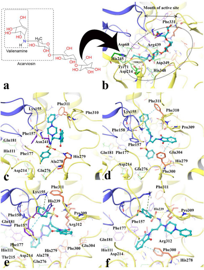
Figure 7.
Acarbose structure (a) and docked representation of acarbose (b), the most active synthesized compounds 4m (c) and 4i (d) and the lowest active compounds 4l (e) and 4n (f) over the α-glucosidase active site. Domain A and B are colored in yellow and blue, respectively. The docked compounds colored in cyan. The α-glucosidase subsides residues include region − 1 and + 1 are in green color also the + 2 and + 3 subsides are in purple and orange, respectively (Maestro Molecular Modeling platform (version 12.5)).

