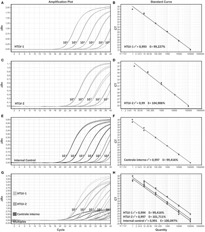
Figure 1.
Amplification plots and standard curves of singleplex and multiplex assays. Singleplex format is demonstrated for (A,B) HTLV-1, (C,D) HTLV-2, and (E,F) internal control, beta-globin. (G,H) Multiplex format for the three targets HTLV-1, HTLV-2, and beta globin. Serial decimal dilutions ranging from 105 to 100 copies/reaction were used in the qPCR reactions. DNA from MT-2 and Gu cell lines were used as positive samples for HTLV-1 and HTLV-2, respectively.

