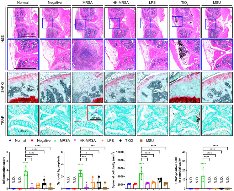
Figure 2.
MRSA Septic arthritis induced inflammation, destruction of articular cartilage, and osteolysis. C57BL/6 mice received intraarticular knee injections of either MRSA (1×10⁵ CFU/10 μL), HK-MRSA (1×10⁵ CFU/10 μL), LPS (200 µg/mL), TiO2 (3 mg/mL), or MSU (200 µg/mL) with select mice receiving DPBS injection to serve as a negative control (n = 4 per group). Ten days after inoculation, mice were sacrificed, and tissues obtained for histological analyses. Paraffin-embedded knee joint tissues were sectioned and histologically stained with H&E, SAF O, or TRAP (scale bars: 2,000, 1,000, and 500 μm). The inflammation score, synovial hyperplasia, synovial cellularity, and TRAP-positive cells were measured using H&E- and TRAP-stained images. Error bars show means ± SD. One-way ANOVA with Tukey’s post hoc analysis was used to assess statistical significance when compared to the negative control group or MRSA group (*p < 0.05 or **p < 0.01 or ***p < 0.001 or ****p < 0.0001; N.D., not detected).

