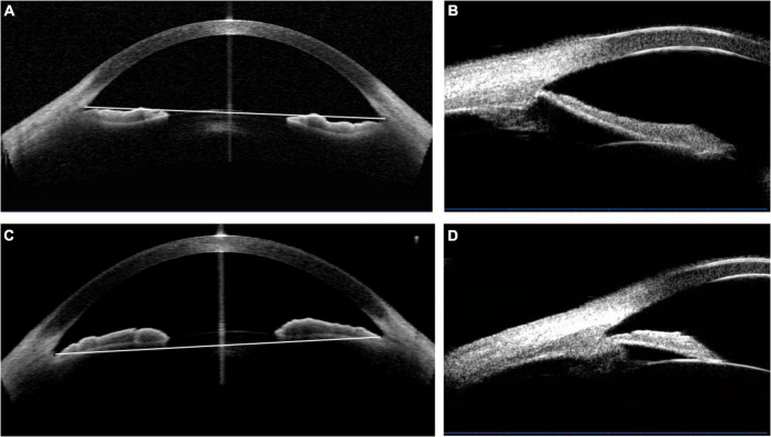
FIGURE 2.
The determination of the concave and convex iris shape based on the anterior-segment optical coherence tomography (AS-OCT) or ultrasound biomicroscopy (UBM) images. (A,B) Concave shape iris: most part of iris locating behind angle-to-angle (ATA) with a concave shape of the iris pigment epithelium, referring to a “bowing” away from the cornea. And a wide sulcus could be detected in (B). (C,D) Convex shape iris: most part of iris locating before ATA with a convex shape of the iris pigment epithelium, referring to that the mid-peripheral iris pigment epithelium is “bowed” toward the cornea. Besides, convex shape iris with anteriorly positioned ciliary body was demonstrated in (D).

