
Figure 2.
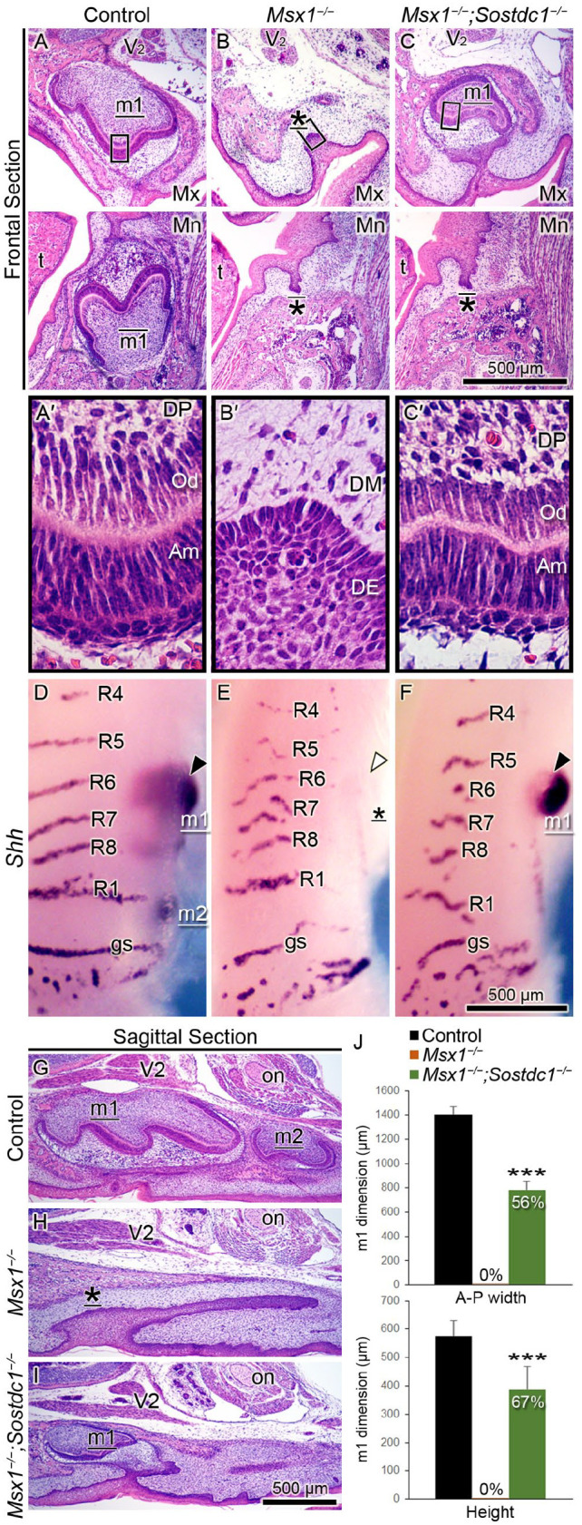
Sostdc1 deletion partially rescues tooth development in Msx1−/− maxillary first molars. (A–C; A′–C′) Hematoxylin and eosin–stained molar tooth germs are shown in frontal sections through the molar tooth germs at birth (P0). (A–C) White horizontal lines divide the maxilla (Mx) and mandible (Mn); the left side is palatal/lingual and the right side is buccal. (A′–C′) High-magnification images of corresponding boxed areas in panels A–C show the histodifferentiation of the ameloblasts (Am), odontoblasts (Od), and dental papilla (DP), as well as the underdifferentiated dental epithelium (DE) and dental mesenchyme (DM). (D–F) Whole-mount visualization of tooth germs in the Mx jaw at embryonic day 16.5 by analyzing Shh mRNA detection, where upper is anterior and lower is posterior. Black and white arrowheads mark Shh-positive and Shh-negative first molar tooth germs, respectively. (G–I) Hematoxylin and eosin–stained molar tooth germs at birth are shown in sagittal sections, where left is anterior and right is posterior. Landmarks used for identification of the tooth germs are the maxillary nerve (V2), which is the second branch of the trigeminal nerve, the fifth cranial nerve, and the optic nerve (on). Tooth germ is labeled m1 (first molar), m2 (second molar), or * (for developmentally arrested tooth bud). These labels are underlined (for Mx) or overlined (for Mn). t, tongue; R, palatal ruga; gs, “geschmacksstreifen” (taste stripes). Scale bars, 500 µm. n = 8 for each of panels A–C; n = 6 for each of panels D–F; n = 10 for each of panels G–I. (J) Measurements of the anteroposterior (A-P) width and height of the tooth germ, showing reduction by 44% in A-P width and by 33% in height. Results are expressed as mean ± SD. n = 10 for each group. Student t test. ***P ≤ 0.001.