
Figure 3.
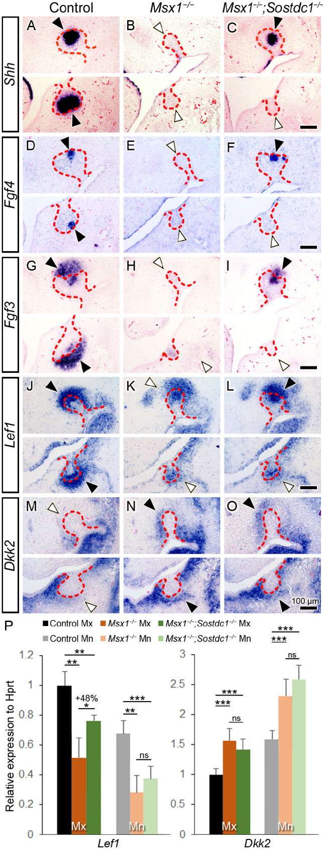
Sostdc1 deletion partially restores Wnt signaling and bud-to-cap transition in Msx1−/− maxillary molars. (A–O) Molecular marker assay in control, Msx1−/−, and Msx1−/−;Sostdc1−/− tooth development at around the bud-to-cap transition (embryonic day 13.75 [E13.75] to E14.25). Primary enamel knot markers Shh (A–C) and Fgf4 (D–F) and a marker for primary enamel knot and dental mesenchyme, Fgf3 (G–I), at the early cap stage (at E14.25). (J–L) Wnt signaling marker Lef1 at bud-to-cap transition (at E14.0). (M–O) Secreted Wnt inhibitor Dkk2 at the late bud stage (at E13.75). In each panel, a white horizontal line divides the maxillary (Mx) and mandibular (Mn) molars; the left side is palatal/lingual and the right side is buccal. The distal end of a tooth germ is marked with an arrowhead: black, stronger mRNA expression; white, weaker. The red dashed line marks the boundary between the dental epithelium and mesenchyme. Scale bars, 100 µm. n = 4 for panels A–O. (P) Real-time quantitative polymerase chain reaction of Lef1 and Dkk2 expression in Mx and Mn molar tooth germs at bud-to-cap transition (at E14.0). Results are expressed as fold change ± SD relative to control Mx. n = 3–5 for Lef1 expression; n = 4–6 for Dkk2 expression. Student t test. *P ≤ 0.05. **P ≤ 0.01. ***P ≤ 0.001. ns, not significant.