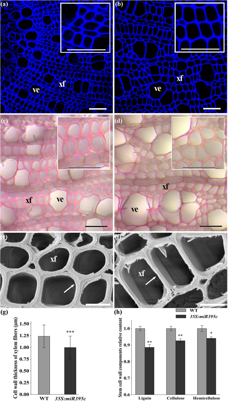
FIGURE 4.
The SCW of the 8th internode of 3-month-old wild type and transgenic poplar. (a) Lignin autofluorescence of wild type. (b) Lignin autofluorescence of miR395c-OE poplar. Phloroglucinol-HCL staining of (c) wild-type and (d) miR395c-OE poplar. Bar, 50 μm. (e) Electron microscopy on the xylem in wild type. (f) Electron microscopy on the xylem in miR395c-OE poplar. The arrow refers to the wall thickness of the xylem fiber. Bar, 10 μm. xf, xylem fiber; ve, vessel. (g) The thickness of fiber cell wall decreased in miR395c-OE poplar. (h) The content of lignin, cellulose, and hemicellulose decreased in miR395c-OE poplar. Insets in (a–d) are high-magnification images of the middle xylem area. Asterisks indicate significant difference between transgenic and WT plants using the Student’s t-test, *p < 0.05; **p < 0.01; ***p < 0.001.

