
Fig. 6.
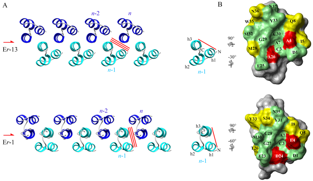
Intra-chain interface conservation between the Er-13 and Er-1 crystal structures. (A) Top view of the Er-13 and Er-1 chains propagating along a two-fold screw symmetry axis (symbol on the left). The red bars indicate the interfaces #1 between one molecule n and the nearest neighbor one, n – 1, that is conserved between the two chains. (B) View of the faces of molecules n – 1 (red bar) formed by helices 1 and 3 and involved in the interface #1. Residues that are conserved, semi-conserved, or not-conserved are colored green, yellow and red, respectively. With respect to their orientation in A, molecules are both first rotated by 90° about the horizontal axis, and then by −30° (Er-13) or −60° (Er-1) about the vertical axis, in order to bring them to the same orientation.