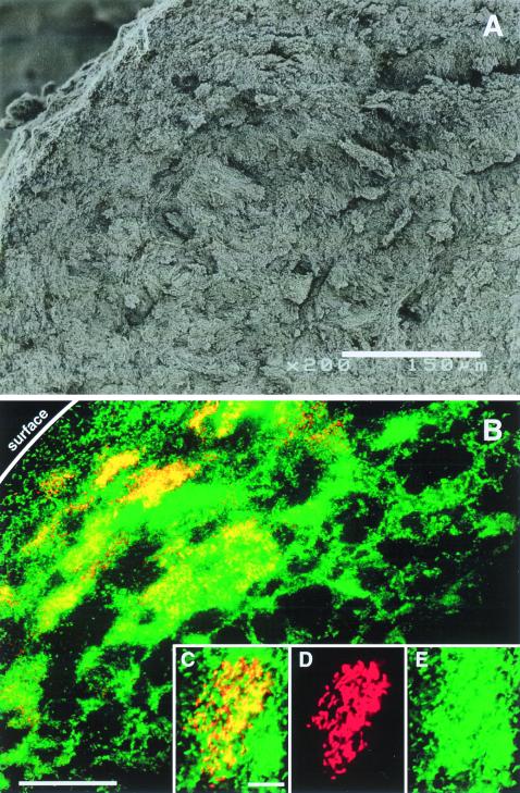
FIG. 5.
Scanning electron micrograph and in situ hybridization of sections from thermophilic granules viewed by confocal laser scanning microscopy. (A) Scanning electron micrograph of a section of a thermophilic sludge granule (bar, 150 μm); (B) section hybridized with the rhodamine-labeled TGP690 probe (red) and the Cy-5-labeled probe MB1174, specific for Methanobacteriaceae (green) (bar, 50 μm); (C to E) magnifications of a microcolony of strain SI and Methanobacterium-like cells (bar, 10 μm): (C) double staining with the TGP690 and MB1174 probes, (D) only signals from the TGP690 probe from the same field as that in panel C, and (E) only signals from the MB1174 probe from the same field as that in panel C.

