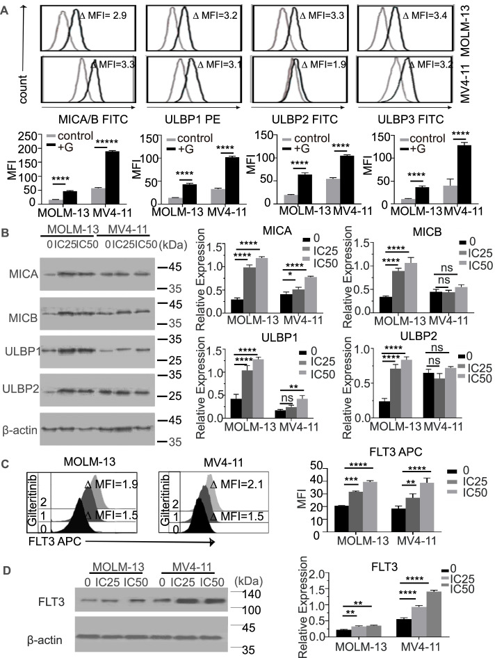
Fig. 4.
Gilteritinib upregulated the expression of NKG2DLs and FLT3 in MOLM-13 and MV4-11 cell lines. A Flow cytometry analysis showed that the expression of NKG2DLs (MICA/B and ULBP1/3) in MOLM-13 and MV4-11 cells was significantly upregulated with gilteritinib treatment. Histograms show NKG2DL expression on MOLM-13 cells and MV4-11 cells in the absence (grey) and presence (black) of IC25-gilteritinib for 24 h. Inset numbers show the ratio in MFI of treated/nontreated cells. B Western blotting (left) showed that the protein levels of all NKG2DLs in the MOLM-13 cell line were significantly increased with gilteritinib treatment, but in the MV4-11 cell line, only the MICA and ULBP1 levels were upregulated. The diagrams (right) show the relative expression as the grey intensity ratio of the NKG2DL (MICA ~ B, ULBP1 ~ 2) protein band to the internal control (C) Flow cytometry analysis of FLT3 expression on cell lines (MOLM-13 and MV4-11) with or without gilteritinib treatment. The histograms show FLT3 expression in MOLM-13 and MV4-11 cells treated without gilteritinib (0), 24-h IC25 gilteritinib (1), and 24-h IC50 gilteritinib (2). D Western blotting (left) indicated that FLT3 expression in both cell lines was significantly upregulated by gilteritinib pre-treatment. The diagrams (right) show the relative expression as the grey intensity ratio of the FLT3 protein band to the internal control. G, gilteritinib; FITC, fluorescein isothiocyanate; MFI, mean fluorescence intensity. ns, not significant. * p < 0.05; ** p < 0.01; *** p < 0.001; **** p < 0.0001

