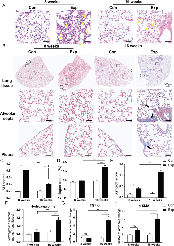
Fig. 2.
Effects of real-ambient PM exposure on chronic lung injury. Whole-slide images of lung sections stained with H&E in control and exposure group following 8-week and 16-week PM exposure. A Representative images of H&E-stained lung sections, displaying the pathological changes in the control and exposure groups following 8-week and 16-week exposure (scale bar = 50 μm). Yellow bold arrows indicate the interstitial neutrophils infiltration (N = 8). B Representative whole-slide images of lung sections stained with H&E in control and exposure group following 8-week and 16-week PM exposure (scale bar = 500 μm) and representative images of Masson's trichrome stained lung sections, showing the collagen deposition in different groups (scale bar = 50 μm). Black thin arrows indicate alveolar septa with gentle fibrotic changes and black bold arrows indicate the fibrotic changes with knot-like formation or pleural plaque formation. C ALI scores were calculated in different groups (N = 8). D Collagen content (%) in lung tissue was calculated as the ratio of labeled blue areas to total area of lung section (%/μm2 total area) upon Masson's trichrome staining (N = 8). E Ashcroft scores were calculated in the groups (N = 8). F Pulmonary hydroxyproline content in the control and exposure groups following 8-week and 16-week exposure (N = 3). G, H The relative mRNA expression levels of TGF-β, and profibrotic factor α-SMA in lung tissue of different groups (N = 5). Photographs were scanned by TissueFAXS analysis system. All magnifications are at 200X. Scale bar = 50 μm. The results were presented as mean ± SD. NS: Not significant; *P < 0.05; **P < 0.01; ***P < 0.001 compared with the control mice. Con: air-filtered control group; Exp: PM exposed group; ALI: acute lung injury

