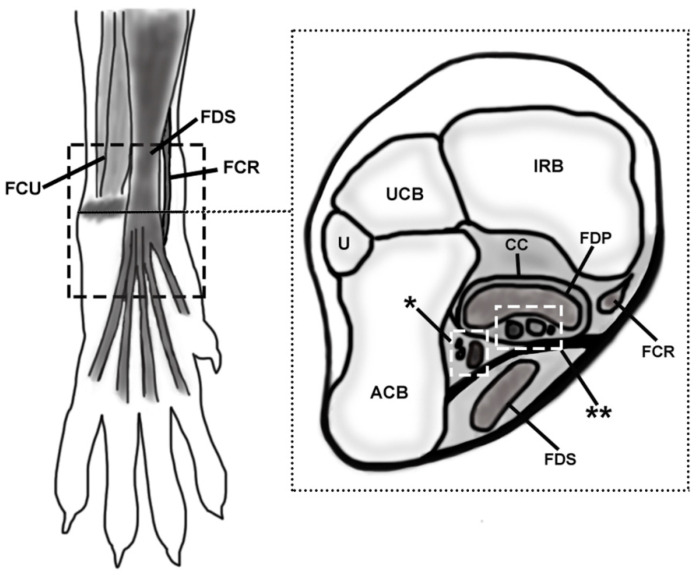
Figure 1.
Schematic representation of the simplified anatomic location of the palmar structures of the canine carpus. The discontinuous lined rectangle in the left image (a palmar view of the carpus) delimitates the studied area. A transverse slice was drawn to show the deeper structures at the level of the accessory carpal bone (represented with a shadow in the left image and as ACB in the right one). The level of the transverse slice is signaled with a black line in the palmar view. FCU, flexor carpi ulnaris; FDS, flexor digitorum superficialis; FCR, flexor carpi radialis; U, ulna; UCB, ulnar carpal bone; IRB, intermedioradial bone; CC, canalis carpi; FDP, flexor digitorum profundus; asterisk, ulnar neurovascular package (arteria, vena, and nervus ulnaris); double asterisk, median neurovascular package (arteria and vena mediana, nervus medianus) and interflexorius muscle.

