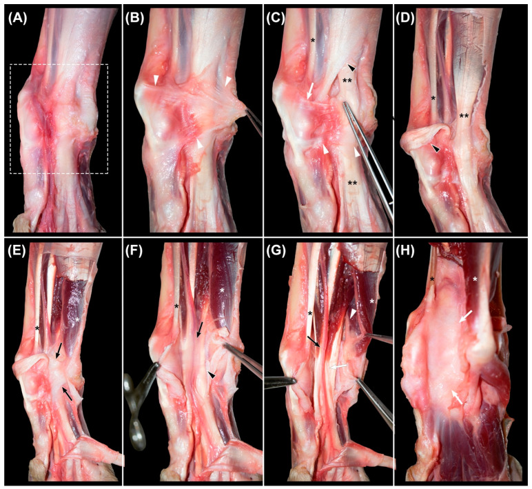
Figure 2.
Sequential images of the dissection of the palmar region of the canine carpus. (A): area under study (rectangle) after removing the skin and subcutaneous fat. (B): identification of the superficial part of the flexor retinaculum (arrowheads). (C): dissected proximal (white arrow) and distal (white arrowheads) bands of the superficial part of the flexor retinaculum and the distal attachment of the fascia of the antebrachium (black arrowhead), and (D): aforementioned structures reclined. Double asterisk (C,D): tendon of the flexor digitorum communis muscle. (E): identification of the deep part of the flexor retinaculum (black arrows) and (F): its incision to access the canalis carpi. The neurovascular structures and tendons of the interflexorius muscle surrounded by loose connective tissue and a small amount of fat (black arrow), overlying the tendon of the flexor digitorum profundus muscle (black arrowhead). (G): dissection of the arteria and vena mediana and nervus medianus (black arrow) and identification of the tendons of the interflexorius muscle (white arrow). The nervus ulnaris (white arrowhead), lateral to the canalis carpi and deep to the flexor carpi ulnaris muscle (white asterisk). (H): after the removal of the structures of the canalis carpi, the underlying palmar common ligament is seen as a smooth surface (white arrows). Black asterisk (C–H): tendon of the flexor carpi radialis muscle.

