
Figure 3.
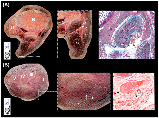
(A,B): transverse slices of the canine carpus. (A): at the distal part of the radius (R) and ulna (U), with magnification of the canalis carpi and complementary histologic section stained with OMSB. (B): at the level of the carpal bones (I–V) with magnification of the canalis carpi and complementary histologic section stained with Picro Sirius Red. The canalis carpi are outlined with dots (rectangle), and its components are shown in the magnified image and in the stained sections. The Picro Sirius Red stained section shows the collagen fibers that separate the structures of the canalis carpi from the nervus ulnaris (black asterisk) and the flexor digitorum superficialis muscle (white asterisk). Black arrows: flexor digitorum profundus muscle; black arrowheads: interflexorius muscle; white arrows: arteria mediana; white arrowheads: nervus medianus; double white asterisk: flexor carpi ulnaris muscle. In the left corner below the transverse slices, a scheme represents their corresponding level at the carpus.