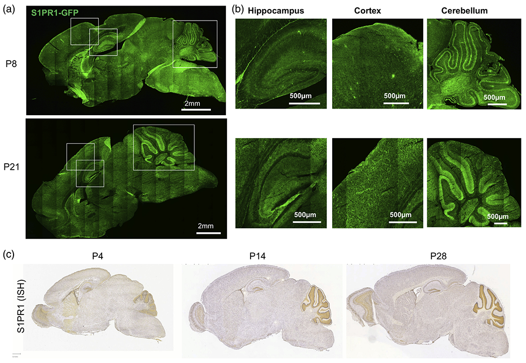
FIGURE 2.

Widespread expression of S1PR1 in the brain. (a) eGFP fluorescence of sagittal sections of S1PR1-eGFP mouse brains at P8 (top) and P21 (bottom). (b) Enlarged images of the indicated cortical, hippocampal and cerebellar regions from panel (a). (c) In situ hybridization (ISH) data for S1PR1 (Allen brain atlas) at the indicated time points of mouse brain development
