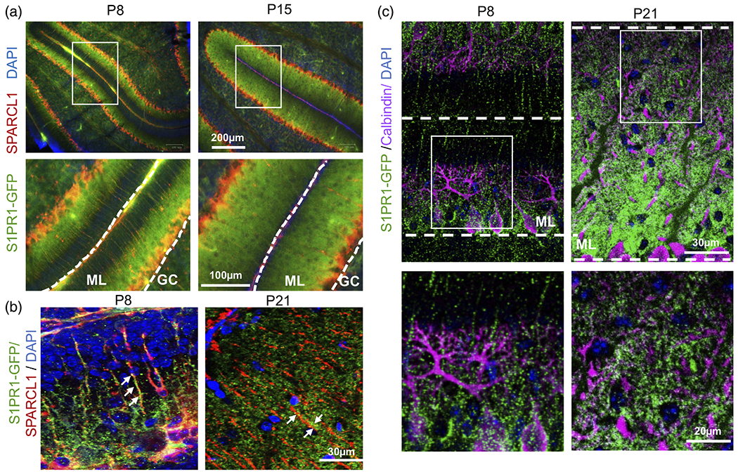
FIGURE 5.

Developmental expression of S1PR1 by Bregman glia. (a–c) Confocal images of S1PR1-eGFP (green) of cerebellar sections at postnatal days P8, P15, and P21 from S1PR1-eGFP transgenic mice immunostained for SPARCL1 (red) or calbindin (magenta). Enlarged boxed areas are shown. GC, granular layer; ML, molecular layer
