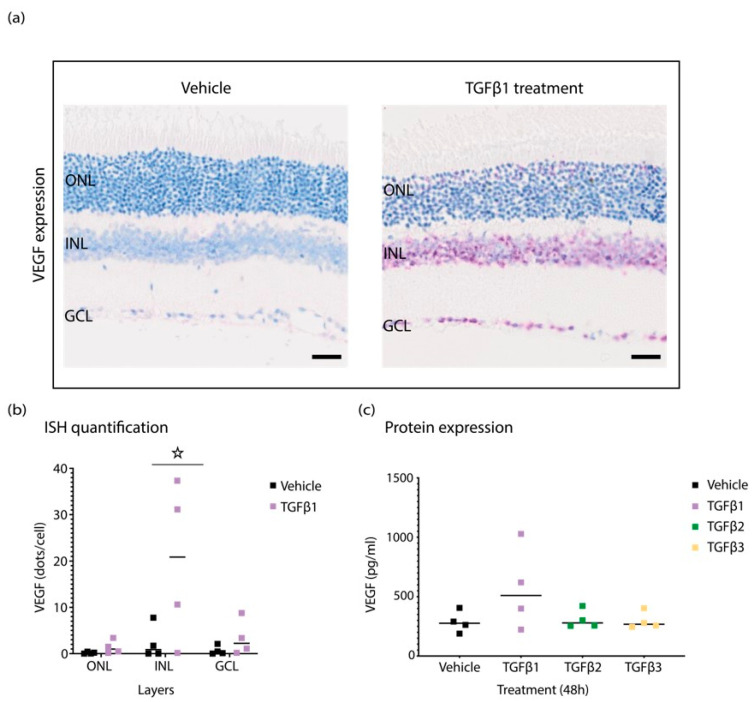
Figure 3.
Retinas from TGFβ1-injected eyes show increased levels of VEGF expression. (a) Representative ISH staining shows elevated levels of VEGF in TGFβ1-treated retina in comparison to vehicle-injected controls. (b) Quantification of ISH staining with HALO. (c) The analysis of cytokine levels with Luminex shows increase in VEGF expression at the protein level in ocular fluid of TGFβ1-injected eyes. Graphs show mean ± SD. All experiments were performed with four replicates, and asterisks represent p < 0.05.

