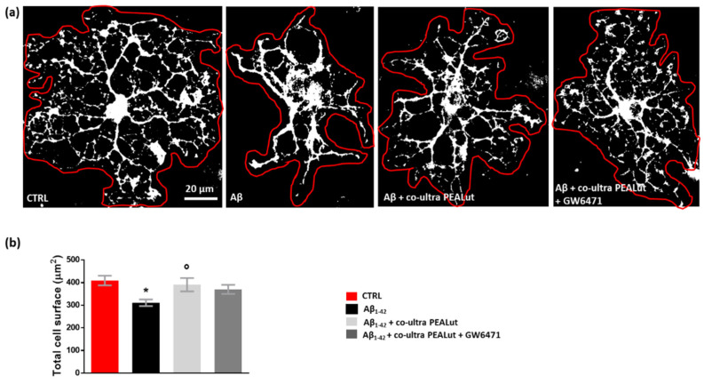
Figure 5.
Co-ultra PEALut prevents oligodendrocyte shrinkage caused by Aβ1–42. (a) Representative thresholded (white color) photomicrographs of MBP+ cells taken under a 40× objective mounted on a fluorescent microscope. Scale bar is 20 μm. (b) Quantification of total MBP+ cell surface area expressed in µm2. Cell contours were manually drawn using Fiji. Data are plotted as mean ± SEM. * p < 0.05 vs. CTRL; ° p < 0.05 vs. Aβ (SNK test).

