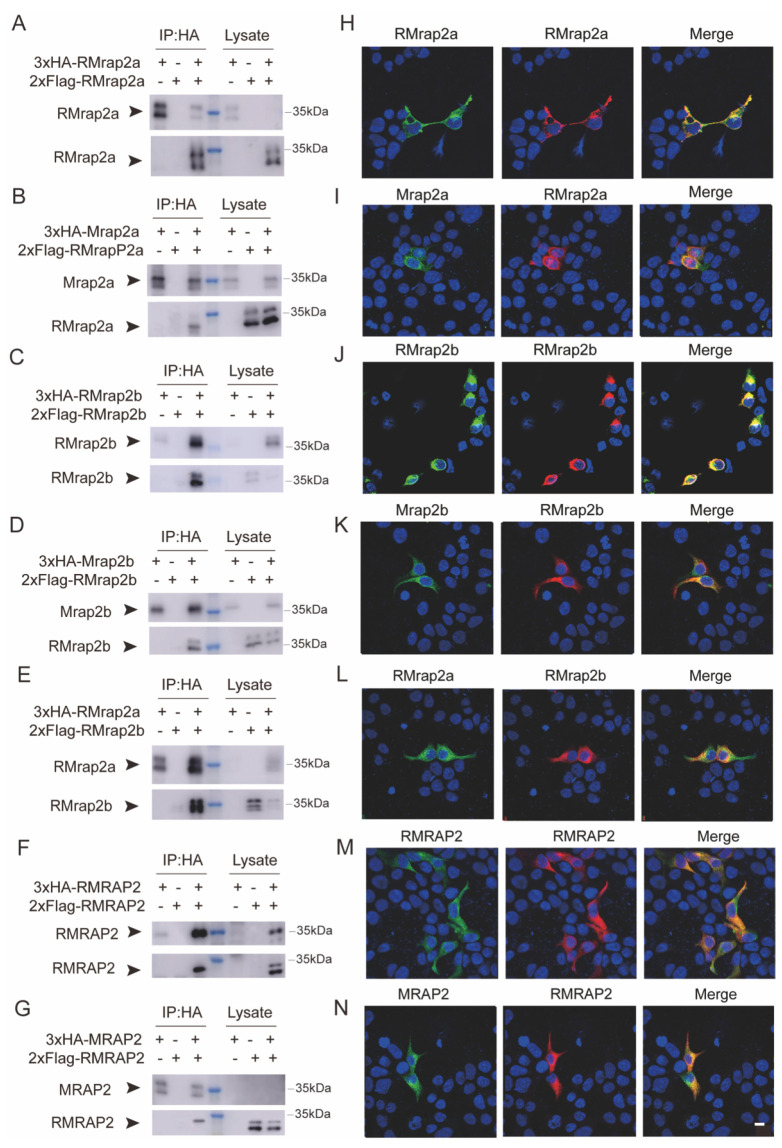
Figure 2.
Formation of homodimers and heterodimers of RMRAP2 and RMrap2a/b. (A,B) The dimerization of RMrap2a with itself (A) or with Mrap2a (B). The blue marker indicates approximate Mol. wt. of RMrap2a and Mrap2a. (C,D) The dimerization of RMrap2b with itself (C) or with Mrap2b (D). (E) Co-immunofluorescence of RMrap2a and RMrap2b. (F,G) The dimerization of mouse RMRAP2 with itself (J) or with MRAP2 (K). (H–N) Immunofluorescence of the co-localization of the CoIP protein complexes on the plasma membrane. Scale bar = 10 μm. The uncropped Western blot figures were presented in Figure S2.

