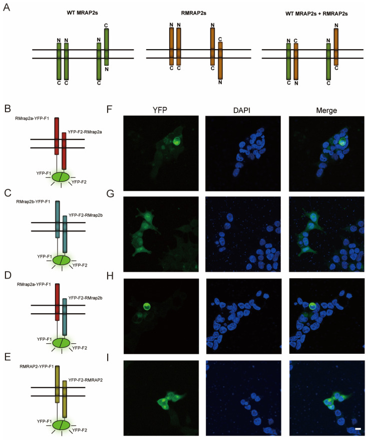
Figure 3.
RMrap2a/b and RMRAP2 form antiparallel dimers on the plasma membrane. (A) Schematic illustration of parallel and antiparallel dimers of WT MRAP2s (left), RMRAP2s (middle) as well as WT MRAP2 and RMRAP2 dimers (right). (B–E) The schematic diagrams illustrate the principle of YFP fluorescence emission and the localization of YFP-F1/F2 on the fused protein. Red: RMrap2a, blue: RMrap2b, yellow: RMRAP2. (F–I) YFP fluorescent and DAPI under confocal microscope. Scale bar = 10 μm.

