Figure 9.
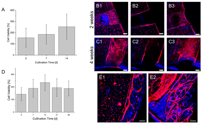
Co-Cultures of HUVEC and HepG2 (A–C) and HUVEC, HepG2 and fibroblasts (D,E) in a prevascularized construct. (A): Cell viability of the co-culture within a culture time of 2 weeks. (B): Endothelialization of the hollow strand by HUVEC in the co-culture after 2 weeks. (C): Endothelialization of the hollow strand by HUVEC in the co-culture after 4 weeks. Sections 1–3: Maximum intensity projection of different segments of the hollow strands. 1: lower area; 2: middle area; 3: upper area. A scheme of the visualized areas can be seen in the Supplementary Materials. (D): Cell viability of the triplet co-culture within a culture time of 4 weeks. The cell viability of each day is normalized to the signal on day 1 after bioprinting (also in A). (E1): Formation of vascular structures by HUVEC in the co-culture after 2 weeks. (E2): Formation of vascular structures by HUVEC in the co-culture after 4 weeks. Blue: DAPI; Red: CD 31; Scale bar: 100 µm.

